Overview

Over the years the VCU Department of Radiology has evolved to meet the needs of our faculty, trainees and patients. Our organizational framework consists of four divisions with ten subspecialty sections and more than 45 faculty radiologists and nuclear medicine physicians. Our framework facilitates collaboration and helps us achieve our mission to be an Image of Excellence.

Our divisions and subspecialty sections enhance our strategic decision-making capabilities and advanced-specialized training. Our framework also improves our ability to identify, recruit and promote the most talented faculty so we can continually meet the requirements of an ever-changing health care environment.

Expertise ranges from interventional and non-interventional radiology to nuclear medicine and neuroradiology with interpretation of computed tomography, magnetic resonance imaging, and ultrasound. Our faculty and staff provide a full range of radiology services with the utmost patient care and clinical excellence in an academic medical institution.


Diagnostic Radiology Sections

Diagnostic Radiology includes several special radiology sections

Abdominal Imaging Section

Abdominal Imaging uses imaging and interventions to diagnose and treat conditions of the abdominal and pelvic regions.

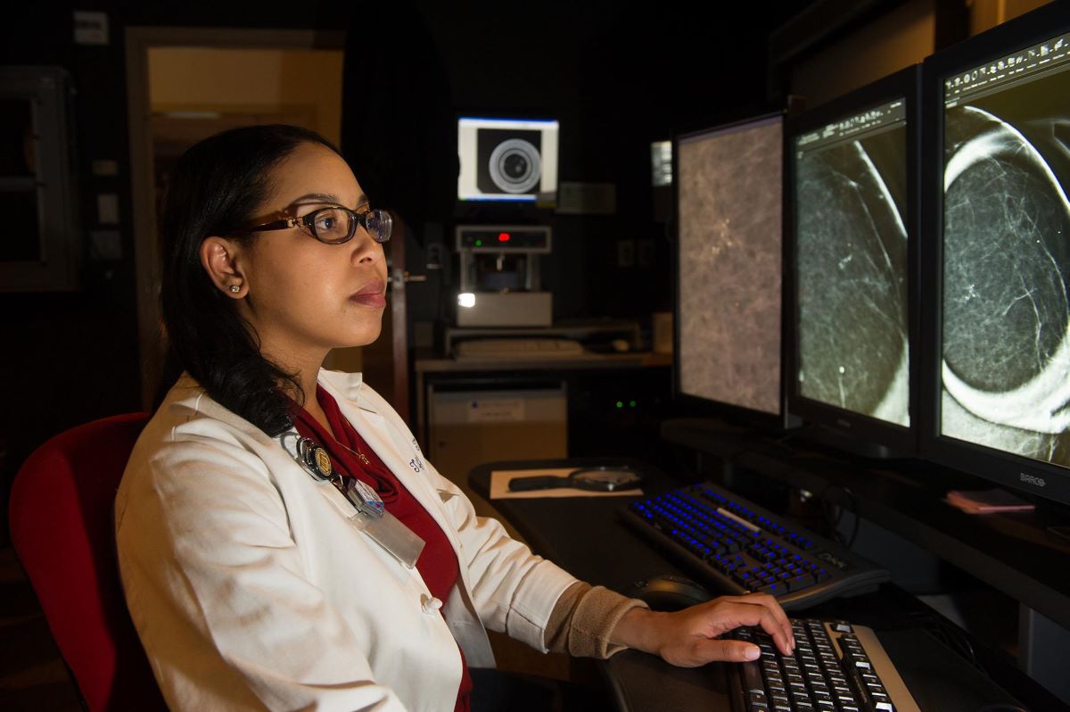
Breast Imaging Section

Breast Imaging provides patients with personalized, high-quality breast health evaluations.

Cardiothoracic Imaging Section

Cardiothoracic Imaging specializes in the diagnosis of diseases and disorders of the cardiopulmonary system and mediastinum.

Emergency Radiology Section

Emergency Radiology interprets a wide range of radiology exams performed through the Emergency Department.

Musculoskeletal Imaging and Intervention Section

Musculoskeletal Imaging and Intervention focuses on diagnosis and minimally invasive treatment of the skeleton, ligaments, muscles and joints.

Neuroradiology Section

Neuroradiology provides neuroimaging techniques to diagnose abnormalities of the head, brain, neck and spine.

Pediatric Imaging Section

Pediatric Imaging interprets the best diagnostic imaging studies on children while using the lowest level of radiation.

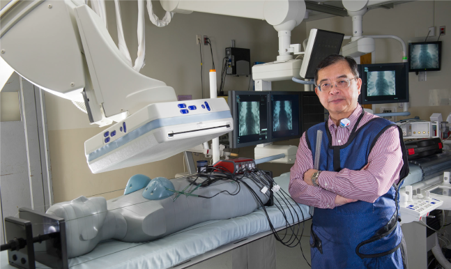
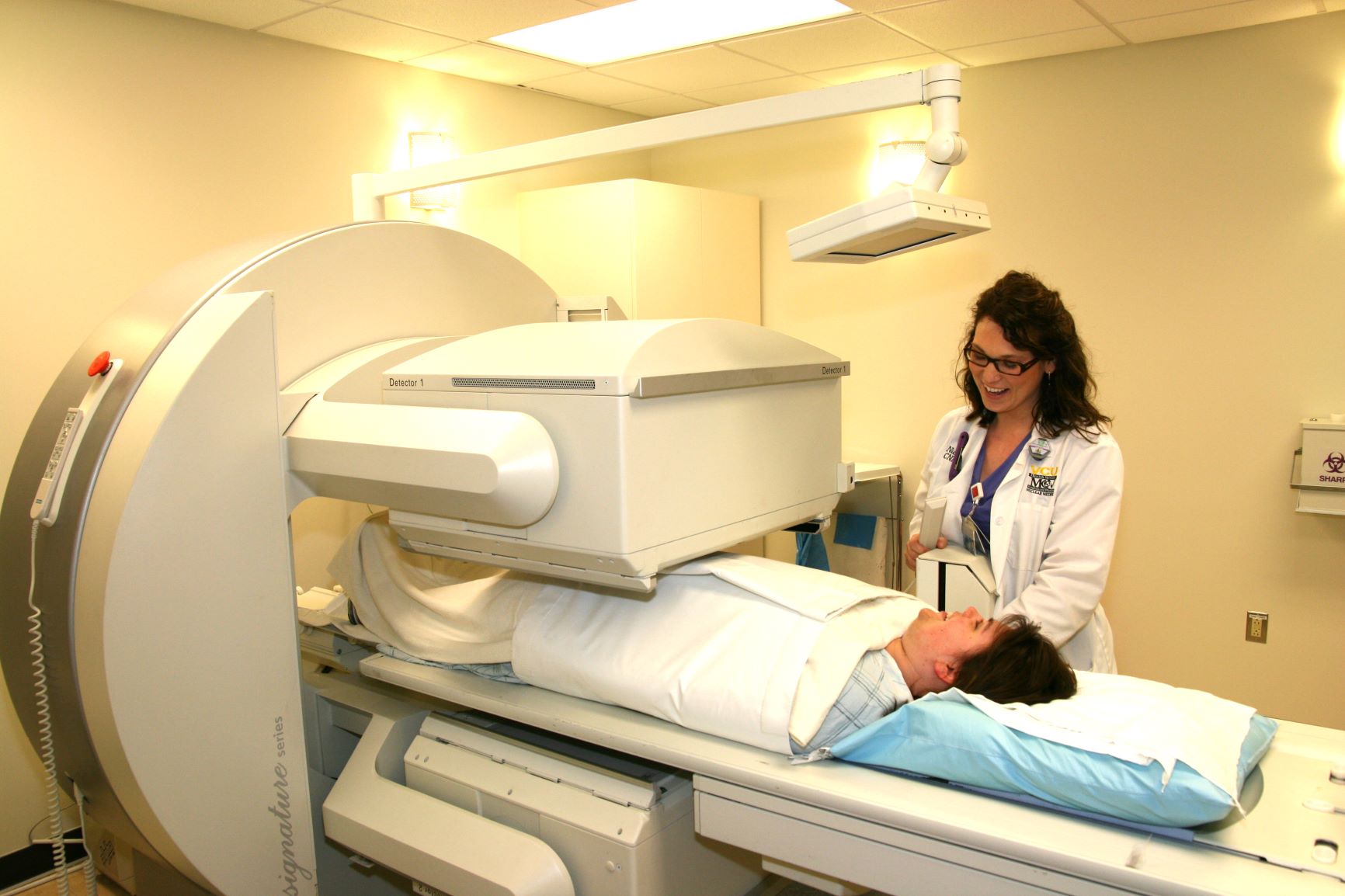
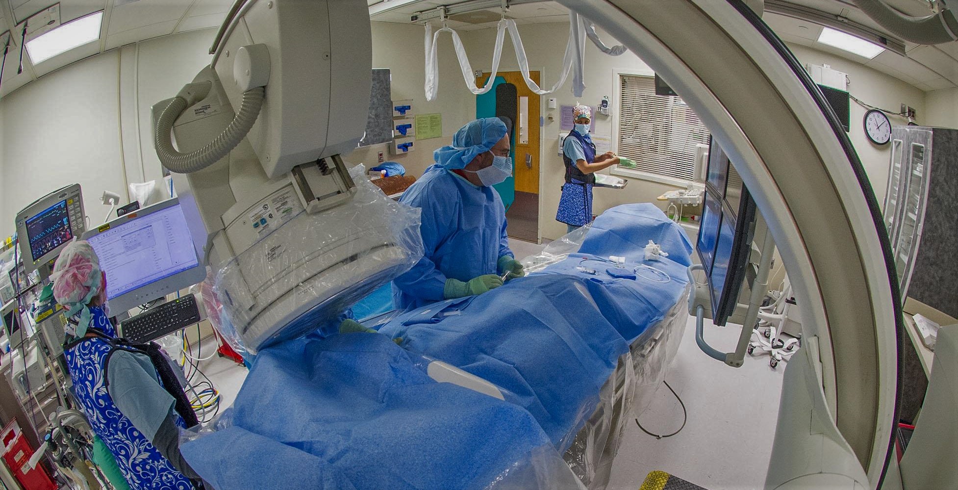
Special Radiology Procedures

Each slide features a different procedure, which emphasizes the expertise of our radiologists.

Advanced Prostate Imaging

Advanced prostate imaging is performed through magnetic resonance imaging (MRI), which uses powerful magnets and radio waves to create detailed images of the prostate. Appropriate treatment recommendations for prostate cancer are based on knowing the accurate staging of the tumor.  During a non-invasive examination, specialists perform multiple advanced imaging techniques, which when evaluated together, can with a high-degree of accuracy detect and locate prostate cancer.

Learn About Advanced Prostate Imaging

Aortic Stent Grafting

The VCU Department of Radiology provides expertise in aortic stent grafting using guided imaging techniques to treat aortic aneurysms. A stent graft is a small tube made of fabric supported by a metal mesh. The stent is typically put in place using a catheter and is designed to seal tightly with the artery above and below the aneurysm. The stent reinforces the wall of the vessel making it safe for blood to flow through. 

Learn About Aortic Stent Grafting

Breast MRI

Breast magnetic resonance imaging (breast MRI) uses a magnetic field to generate images of the breast before and after a contrast injection. The VCU Department of Radiology's Breast Imaging Center was the first facility in Virginia with breast MRI to earn the American College of Radiology Breast Imaging Center of Excellence designation.

Learn About Breast MRI

Computed Tomography Enterography and Magnetic Resonance Enterography

Specialty-trained gastrointestinal radiologists offer computed tomography enterography and magnetic resonance enterography to evaluate the small intestine. These procedures use the cross-sectional imaging technology of computed tomography or magnetic resonance to assess the small intestine, the surrounding mesentery, vasculature and other adjacent structures.

Learn About CTE and MRE

Fetal MRI

Fetal magnetic resonance imaging (fetal MRI) is an imaging technique that provides information about the anatomic structure of a fetus. It particularly excels in the evaluation of the fetal brain and spine. Fetal MRI can be used to help prognosticate outcome in cases of congenital diaphragmatic hernia and fetal lung masses. 

Learn About Fetal MRI

Lung Cancer Screening

Our lung cancer screening program is the first facility in the Commonwealth of Virginia to be designated by the American College of Radiology (ACR) as a Lung Cancer Screening Center. As an ACR designated Lung Cancer Screening facility, the VCU Department of Radiology is dedicated to providing high quality screening care and patient safety.

Learn About Lung Cancer Screening

Peripheral Arterial Disease Treatment

Peripheral arterial disease (PAD) is caused by the buildup of plaque in the arteries of the legs. Plaque is a substance made up of cholesterol, calcium and fibrous tissue.

Our specialty-trained radiologists use guided imaging techniques to perform minimally invasive procedures, including angioplasty and stents, to widen the artery, thereby allowing more blood flow. 

Learn About PAD Treatment

Varicose Veins and Spider Veins Therapy

Interventional radiologists and vascular surgeons collaborate to treat varicose veins and spider veins using advanced radiology techniques and the latest technology.

Learn About Vein Therapies

Uterine Fibroid Embolization

Uterine fibroid embolization (UFE) is a minimally invasive procedure where experienced interventional radiologists use X-ray imaging to guide a catheter placed in the upper thigh to the uterine arteries. Tiny particles are then injected through the catheter into the blood vessels, blocking the blood supply feeding the fibroids, which causes them to shrink and die. UFE is an alternative to traditional treatments that include the surgical removal of the fibroids (myomectomy) or removal of the entire uterus (hysterectomy).

Learn About Uterine Fibroid Embolization

Ultrasound Imaging

Ultrasound is the use of high frequency sound waves to create an accurate image of the inside of the body. Ultrasound generally utilizes a small, portable device, which uses sound waves instead of ionizing radiation.

Learn About Ultrasound

Virtual Colonoscopy (CT Colonography)

The VCU Department of Radiology was among the first in Virginia to offer virtual colonoscopy, which is medically known as computed tomography (CT) colonography. The advanced imaging procedure is non-invasive and allows an evaluation of the entire colon, without the risks, such as bowel perforation, or complications from sedation associated with standard colonography. 

Learn About CT Colonography